DICOM XA / RF playback
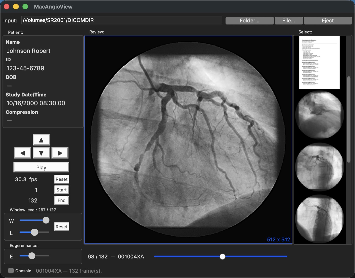
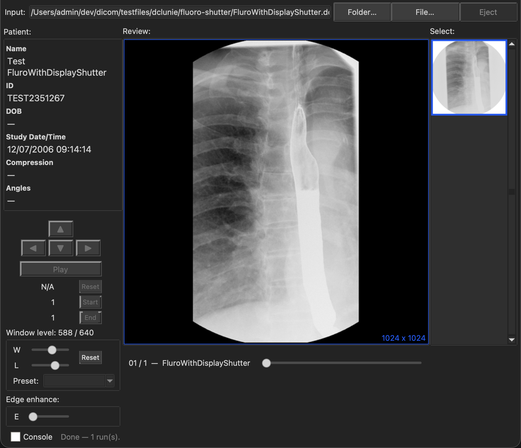
Open and play multi-frame DICOM XA cine-angiogram and radiofluoroscopic (RF) runs, including lossless-JPEG compressed studies from cardiac catheter X-ray systems.
Multiframe ultrasound (US)
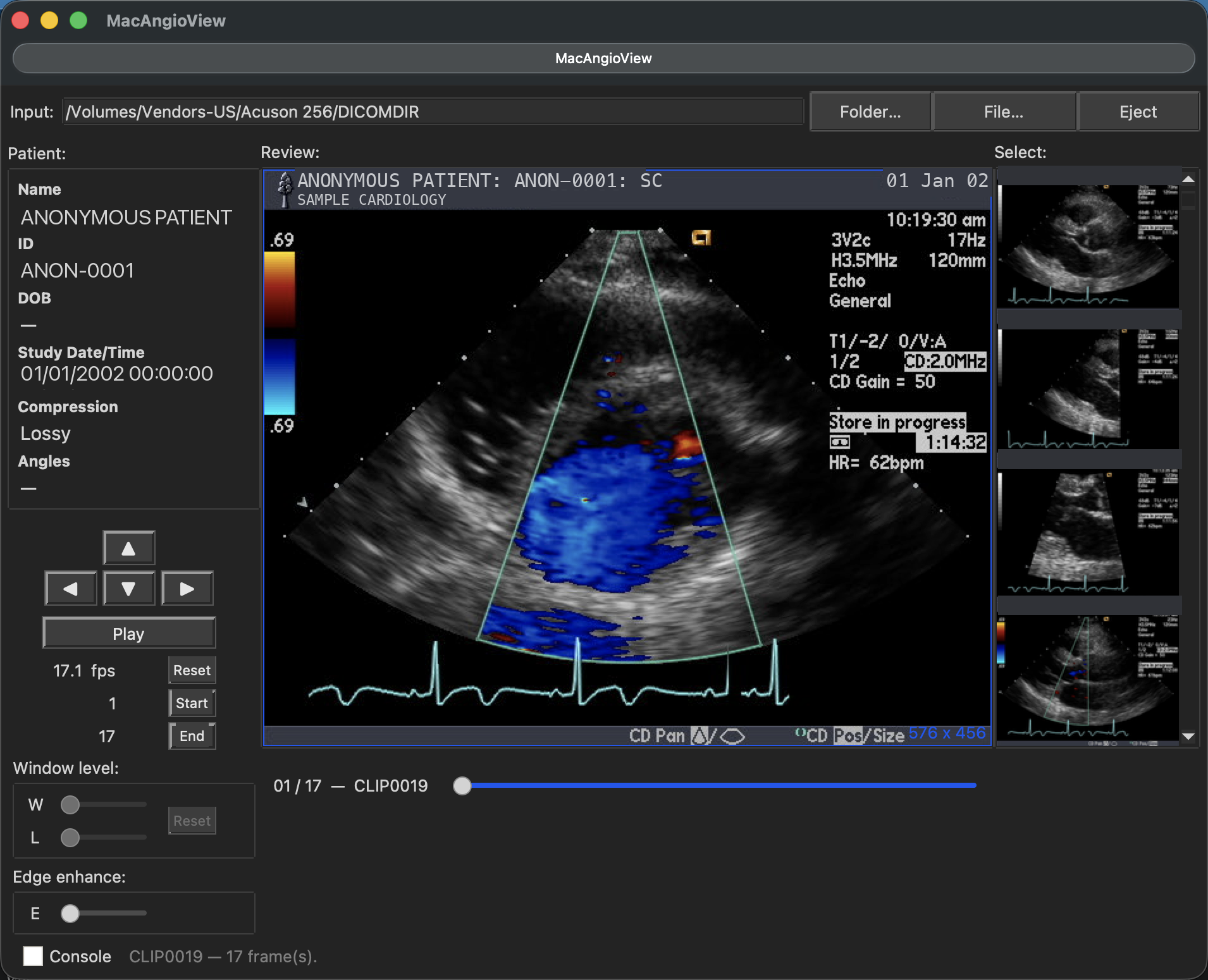
Play back multiframe DICOM ultrasound cine loops — review echo and vascular ultrasound studies alongside your angiograms.
CT/CTA series
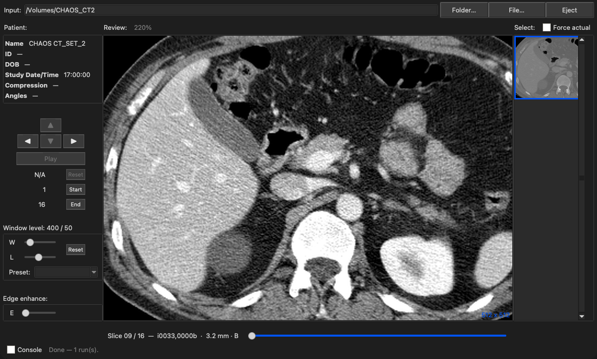
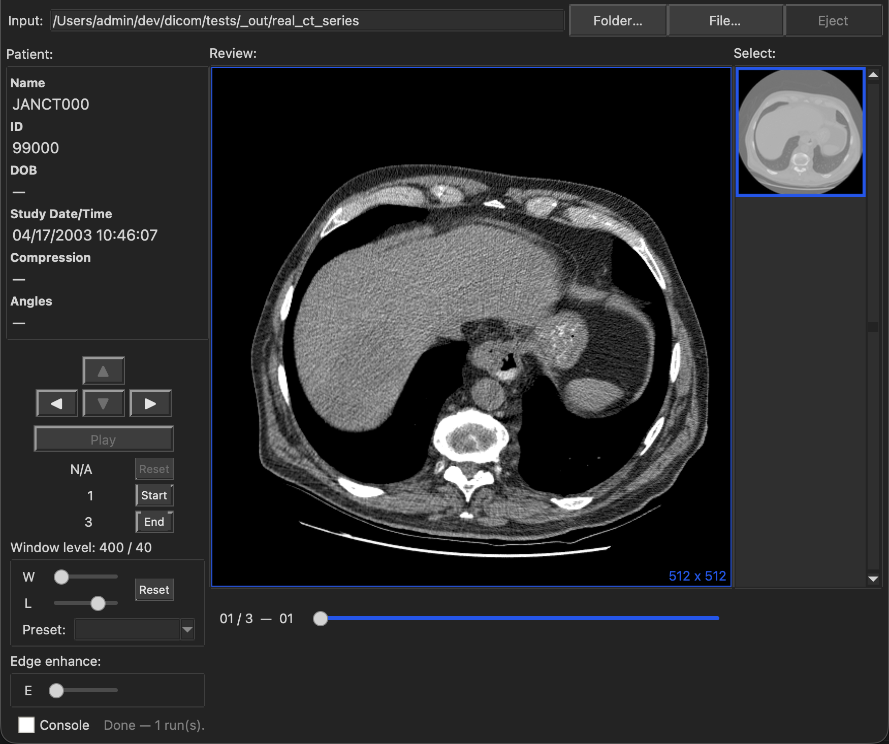
A study's CT slices are grouped into one browsable stack, ordered by their true position in the patient rather than by file numbering, and stepped slice by slice. A series holding several reconstructions or cardiac phases opens as one stack each, with a Preset picker for multi-valued window/level (soft tissue, bone, lung).
MR series
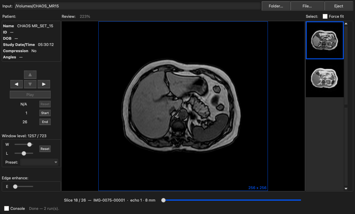
MR studies are read alongside the rest of a patient's imaging, grouped into browsable stacks in anatomical order. A sequence acquired at more than one echo opens as one stack per echo, so each echo can be reviewed on its own rather than interleaved.
Broad system compatibility
Reads studies exported from many cath-lab, ultrasound, and CT imaging systems — load a study CD or exported folder and browse the runs.
Frame-by-frame review
Step through frames, adjust playback speed, and review runs at your own pace. Jump straight to any run by name from the Select menu.
DSA subtraction
View angiograms with the standing anatomy subtracted away — automatically when the study provides a mask, or pick any pre-contrast frame yourself and nudge out patient motion.
Export
Export cine runs to standard movie formats for presentations and teaching files. Exports and copied frames match what you see: zoom in and the export is the magnified view, trimmed to any region you set.
Reports & waveforms
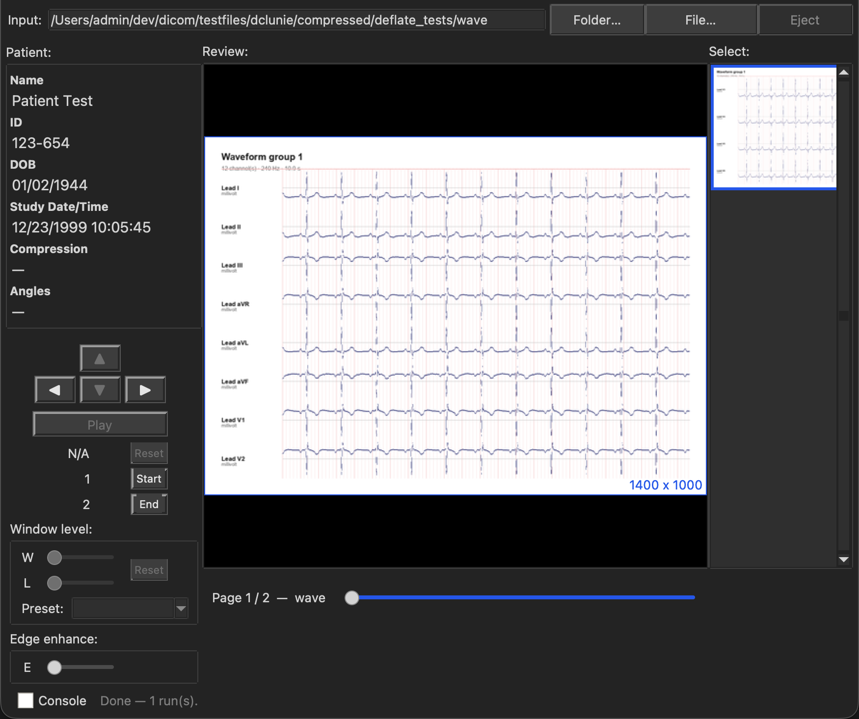
View a study's reports right next to the imaging — encapsulated PDF reports, DICOM Structured Reports (cath, QCA, hemodynamics, ECG summaries), and Waveform Storage instances (12-lead ECG, hemodynamic, cardiac EP) rendered as calibrated lead-strip pages.
Quiz Mode
A self-study workflow — hides a study's own report until you're ready, then lets you record your own Cath, Echo, or Vascular findings and check them against the real report.
Caliper Tool
Two-click distance measurement in millimeters, calibrated automatically from an ultrasound region or XA beam geometry — or manually from a catheter shaft for the most accurate reading.
Native-resolution viewport
Every study renders at its own pixel resolution in a freely resizable window — drag to roam, scroll to zoom under the cursor, drag a box to magnify a region, double-click to switch between actual size and fit to window.
Presentation States
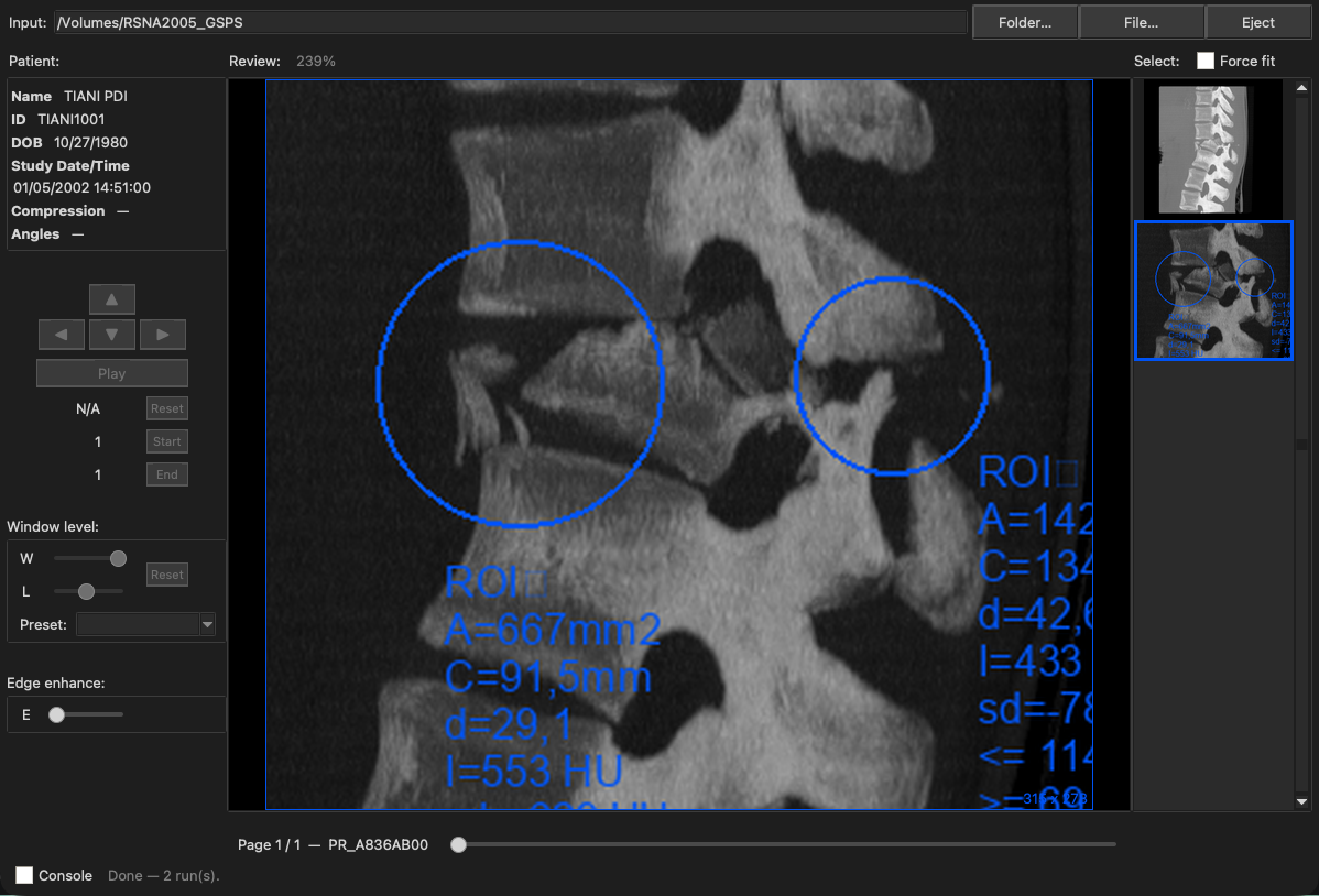
Reads a study's saved annotated views (GSPS): the window, crop, and circle, ellipse, or text markup someone recorded on an image, shown as its own page with the unmarked original still available.
Discs without an index
A folder of DICOM files with no DICOMDIR index opens as one study, so exported folders and discs that lack a catalogue are still readable.